Principles of multivalency-biased affinity selection
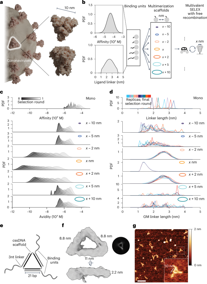
In nature, molecules often coevolve within interconnected, multicomponent assemblies, resulting in the collective development of interfaces. As an analogy, we envisioned the use of a target-tailored multimerization scaffold to evolve synthetic polymers against an oligomeric target, yielding multivalent binders with cooperative binding behaviour within a designed multivalent assembly. Many clinically relevant target proteins exist as multimeric complexes, supporting the application of target-tailored evolution to identify new binders. The evolutionary-conserved trimeric structure of viral capsid glycoprotein complexes is emblematic of many human pathogens such as retroviruses32, coronaviruses33 and orthomyxoviruses34. As a target for the development of our multivalent selection methodology, we opted to use the trimeric SARS-CoV-2 spike protein, arguably one of the most representative members of viral class I fusion proteins (Fig. 1a).
a, Large trimeric glycoprotein complexes are characteristic of many human pathogens as exemplified by SARS-CoV-2 spike protein. Figure created in Blender (https://www.blender.org), using PDB 3JCL as a spike model. b, Schematic for Gillespie simulation of multivalent selection against a homotrimeric target. A random library of binding units with varying spatial tolerances for simultaneous target engagement and a set of multimerization scaffolds with increasing interligand spacing were tested. The initial probability density function (PDF) of affinities (top) and linkers (bottom) for the binding unit library is given. c, Simulation results of multivalent selection for libraries prepared with different multivalent scaffolds show that matching scaffold geometry facilitates the selection of assemblies with the highest avidity. All Gillespie simulations were performed in 10 replicates (n = 10); results are shown as average PDFs. d, The PDF of linkers in the binding unit library in the final round of multivalent selection is determined by the scaffold geometry. GM, geometric mean. e, Schematic representation of MEDUSA’s molecular architecture. f, Cryo-EM 3D class-average electron density of a designed MEDUSA. The respective Fourier shell correlation analysis is provided in Supplementary Fig. 4. g, Representative atomic force microscopy image of MEDUSA. Scale bar, 20 nm.
The performance of multivalent binders relies heavily on the scaffold joining the ligands because this determines their orientation and overall structural flexibility24. Previous modelling results suggest that maximal binding affinity can be achieved when the binder’s core is rigid and matches the target’s size, and the linkers have an average end-to-end distance slightly longer than the distance between the binder’s core and cognate binding epitopes15. Building on these results, we developed a multiscale computational framework for simulating multivalent SELEX (Supplementary Fig. 1) through modelling stochastic selection dynamics35. Simulations used a fixed triangular target with dimensions derived from the spike protein (marked as x) to examine selections conducted with various scaffold geometries (Fig. 1b) and explored how binding affinities and linker lengths evolved under different spatial constraints.
In the multivalent SELEX process, ligand and linker distributions adapt over successive rounds of selection, yielding the best avidities when the scaffold geometry closely aligns with the target (Fig. 1c). Minor deviations in scaffold size (for example, <x − 2 nm or >x + 2 nm) quickly limit avidities to the micromolar range, demonstrating the critical role of scaffold geometry in enabling effective multivalent binding. Scaffold geometry directly influences linker length selection (Fig. 1d): when the scaffold geometry matches the target geometry, libraries converge toward minimal linker lengths that facilitate multivalent engagement by precise alignment with binding epitopes. In contrast, monovalent libraries exhibit a random distribution of linker lengths after selection because they are not subjected to spatial constraints. Scaffolds deviating slightly from the optimal geometry still support multivalent binding by selecting longer linkers. However, scaffolds outside the spatial tolerance of the ligand library fail to favour multivalent interactions. In these cases, selection reverts to a monovalent-like regime, without selective pressure on linker length. These findings underscore the significance of scaffold geometry in determining spatial constraints, providing predictive design principles for optimizing ligand selection in multivalent systems.
Based on simulation results, we established several design principles for the scaffold and binding unit library. First, the scaffold geometry should closely match the target’s size and shape to maximize the contribution of avidity. Second, for matching scaffolds, linkers should be of minimal length to not disrupt the advantages of a geometric fit. We explored several potential scaffold structures that would satisfy these criteria and allow for the display of binding units at ~10 nm pairwise distances within a trivalent framework, reflecting the overall dimensions and rotational symmetry of SARS-CoV-2 spike protein (Supplementary Fig. 2a,b). We settled on a cyclic single-stranded DNA (cssDNA) scaffold due to its nuclease stability, size and ease of preparation (Supplementary Figs. 2c and 3). The architecture of MEDUSA emerges from the three 21-nucleotide binding unit-hybridization regions, separated by 2 T hinges within cssDNA (Fig. 1e–g). This configuration allows the binding units to be positioned at every second turn of the DNA, displaying them on the same face of the scaffold. Following the second design principle, we opted to keep the linkers short, placing 3 nucleotides (~1.6 nm (ref. 36)) between the scaffold-hybridization region and the binding region.
Multivalent assemblies of functionalized nucleic acids
Besides structural programmability, a major advantage of DNA as scaffold is its compatibility with numerous evolvable polymers, such as DNA, RNA9,10, slow off-rate modified aptamers37, highly side-chain-functionalized nucleic acid polymers38, PNA39 and acyclic l-threoninol nucleic acid40. This compatibility makes it possible to construct hybrid nanostructures that merge the versatile design potential of DNA with the chemical diversity of proteins and beyond38,41. We combine the cssDNA scaffold with functionalized nucleic acid polymer (FNAP) binding units, which together create the MEDUSA for affinity evolution of hybrid multivalent targeting nanomaterials. FNAP is a base-modified nucleic acid polymer, produced via DNA-ligase-mediated polymerization of trinucleotide building blocks on the DNA template. In the FNAP library design, we partially sacrifice the density of side-chain functionalization in favour of sequence diversity, allowing the library to sample a broader range of secondary structures of the polymer. Structure–activity relationship studies provide the rationale for this choice because often only few side chains are essential for target binding38. A convenient property of MEDUSA is its excellent nuclease resistance, so critical for applications in biological fluids, ensured by the cyclic scaffold and enzyme-inhibiting nucleotide modifications at the 5′ position42,43.
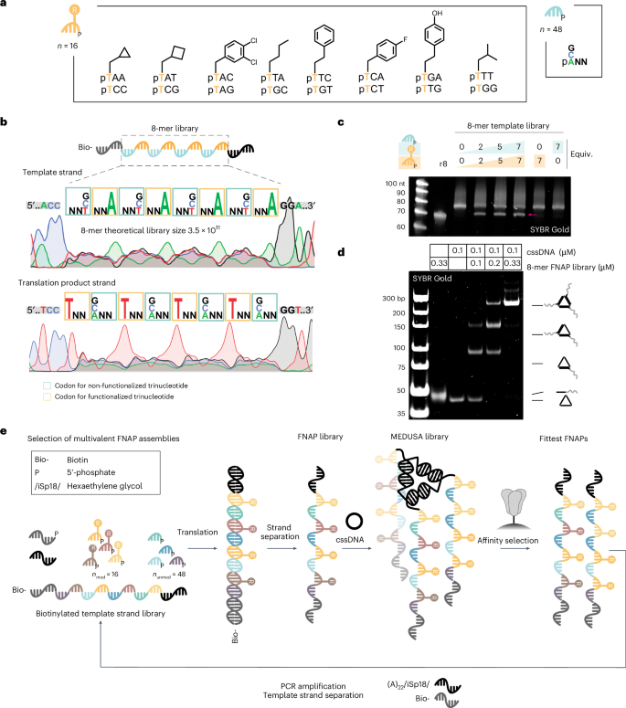
We constructed trinucleotide libraries of 16 side-chain-functionalized trinucleotides encoding 8 different side chains and 48 non-functionalized trinucleotides. Based on previous reports44,45, we limited side-chain diversity to hydrophobic residues, including those that mimic side chains of proteinogenic amino acids and those not found in natural proteins (Fig. 2a, Supplementary Figs. 5 and 6, and Supplementary Table 1). The pertinent template library features an architecture that intersperses low-sequence diversity and side-chain-functionalized trinucleotide regions with high-diversity, non-functionalized trinucleotide regions. The former expands the chemical diversity, while the latter enhances the polymer’s conformational diversity (Fig. 2b). To minimize interference between binding units within MEDUSA, the library length was kept shorter than typical SELEX libraries (40 nt) and the average length of reported spike-binding aptamers (Supplementary Fig. 7a). To estimate the translation efficiency, we prepared corresponding biotinylated template libraries of 8-mer and 12-mer lengths (Supplementary Fig. 7b) and observed a plateau already at 2 equiv. of trinucleotide libraries relative to the template. The maximum translation yield was reached at ~5 equiv., resulting in ~70% for the 8-mer library (Fig. 2c) and ~30% for the 12-mer library, as confirmed by denaturing polyacrylamide gel electrophoresis (PAGE) (Supplementary Fig. 7c,d). Both synthetic FNAP libraries anneal efficiently with the cssDNA scaffold, yielding the trivalent combinatorial MEDUSA library (Fig. 2d, and Supplementary Fig. 7e,f). Due to its higher translation yields and to minimize cross-hybridization between longer binding units, we opted to use the 8-mer library for affinity selections.
a, Structures of functionalized and non-functionalized trinucleotide building blocks for the production of the FNAP library. b, The structure of the 8-mer template library with the corresponding Sanger sequencing electrophoretic traces for direct and reverse sequencing runs. c, Translation reaction of the biotinylated 8-mer template library into the corresponding FNAP library performed at different trinucleotide library concentrations. Non-biotinylated 8-mer library (r8) was loaded as a reference. The FNAP translation product is arrowed. d, Native PAGE analysis of the assembly of MEDUSA using a synthesized FNAP library. e, Scheme of the multivalent selection cycle for trimeric FNAP assemblies.
Each round of selection commences with ligase-mediated translation of the biotinylated template DNA library into a FNAP library using 5′-phosphorylated trinucleotide building blocks. During this process, the ligase catalyses the templated synthesis of the FNAP region between the initiation and termination primers. Following strand separation and purification, the FNAP library is trimerized using the target-tailored cssDNA scaffold and subjected to affinity selection against the target protein, which is immobilized on magnetic beads. For the next selection round, the library is heat eluted and FNAPs are reverse translated back into the DNA template library (Fig. 2e). Due to the target-tailored geometric organization, the selection pressure is directed towards the enrichment of synergistic multivalent binders. Sequences capable of cooperative binding to the target should expand faster due to their slower dissociation rates. With every round, affinity selection based on multivalent interactions becomes more dominant.
Multivalent affinity selections yield a high degree of sequence diversity
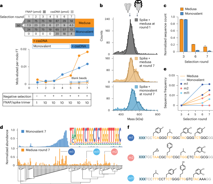
As in SELEX, the frequency of high-affinity sequences in the naïve FNAP library is low, with some binders present in only a single copy. Moreover, the distribution of binding affinities is unknown. To address the high complexity of the naïve library, statistically unfavourable for the stochastic assembly of MEDUSAs during early rounds of selection, two selection strategies were tested: (1) entirely multivalent and (2) a monovalent pre-enrichment during initial rounds, followed by multivalent final selections. We conducted two parallel selection campaigns against the SARS-CoV-2 spike protein (Supplementary Fig. 8). The PAGE-purified starting FNAP library was split and one half (termed ‘monovalent’) first underwent four rounds of monovalent affinity selections before multimerization. The other half was directly subjected to multivalent selections from round 1 (termed ‘medusa’, Fig. 3a, top). The ratio between the FNAP library and target was maintained constant for both strategies throughout the selection campaign (Fig. 3a, bottom). After seven rounds of affinity selection, the binding capacity of both the monovalent and MEDUSA libraries began to plateau, prompting us to conclude the selection campaign (Fig. 3a). The presence of binding sequences in both libraries after selection round 7 was confirmed by mass photometry (Fig. 3b).
a, Progress in spike-binding selection for multivalent (medusa) and monovalently prefocused (monovalent) selection strategies. Bulk affinity of trivalent and monovalent FNAP libraries to trimeric spike protein was assessed by quantifying the amount of FNAPs in the flow-through (FT) versus elution fractions. b, Increase in bulk affinity of trimeric FNAP libraries to spike protein by comparing the MEDUSA assemblies prepared with the FNAP library from selection round 1 versus selection round 7 for both selection strategies using mass photometry. c, Progression of selection process indicated by the decrease in FNAP library complexity using NGS. Data are presented as mean ± s.d. (n = 2, independent sequencing runs). d, Multiple sequence alignment of the top sequences from NGS data for two tested selection strategies with corresponding sequence abundances. The common motif of sequences retrieved from the monovalent selection process is displayed as a nested graph. e, Enrichment of three selected hits over the rounds of selection for two tested selection strategies using NGS. f, Sequences and side-chain structures of selected FNAPs. xxx, scaffold-hybridization region.
Sequencing data at rounds 3, 6 and 7 revealed a steady decrease in the number of unique reads in the sequence pools as selection progressed, indicating the expansion of binding sequences for both selection strategies. The diversity of the final MEDUSA library was around 4.6 times higher than the monovalent library, as estimated from the number of unique sequences in the pool (Fig. 3c and Supplementary Fig. 9a,b). We reason two potential processes could contribute to the difference in the complexities of the libraries: enrichment of synergistically binding sequences that otherwise cannot expand in the monovalent selection regime, and ‘parasitic’ carry-over of low-affinity sequences into the late rounds of selection due to their stochastic incorporation into the MEDUSAs composed of high-affinity sequences. As ‘parasitic’ low-affinity sequences effectively render respective MEDUSAs less active than the assemblies with higher numbers of cooperatively binding sequences, we reason that the selection is directed towards the decrease in the frequencies of low-affinity sequences.
Next-generation sequencing (NGS) data after the final round of selection revealed that the sequence pool for monovalent selection strategy was eventually dominated by one family of FNAPs featuring a common consensus sequence (Fig. 3d, top). Notably, the discovered motif can also be found in several reported DNA aptamers, thereby further validating the specific enrichment of binding sequences46 (Supplementary Fig. 10). In contrast, the sequencing data obtained from the medusa library display a more diverse range of sequences in addition to the aforementioned sequence family (Fig. 3d, bottom). The differences in sequence compositions between the two libraries are also evident in the changes in side-chain frequencies observed across different rounds of selection (Supplementary Fig. 11). To validate the enriched FNAPs, we selected the three most abundant and sequence-unrelated sequences based on the Levenshtein distances matrix (Supplementary Fig. 12). The sequence m1 represents the family of sequences enriched through monovalent selection, while m2 and m11 are the sequences that emerged uniquely through the multivalent, geometry-constrained selection process (Fig. 3e,f).
Evolution with MEDUSA yields unique target selectivity
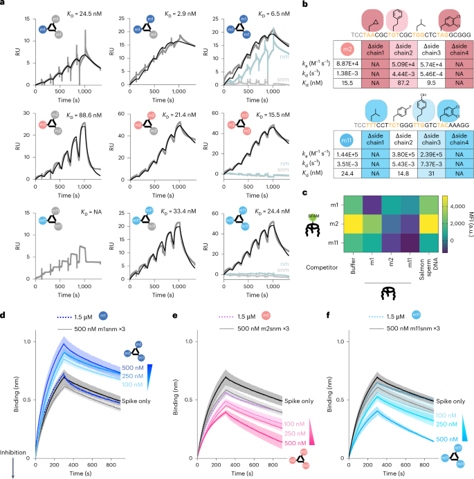
To confirm binding affinity for spike protein, the monovalent binding of the full-length and primer-minimized versions of selected FNAPs (Supplementary Fig. 13a) was tested via surface plasmon resonance (SPR) analysis. The m1 sequence displayed low-nanomolar monovalent affinity for the spike protein (Supplementary Fig. 13b), and m2 showed ~200-nM affinity (Supplementary Fig. 13c). No SPR signal was observed for monovalent m11, even at the highest analyte concentration tested (600 nM). We found that the primer region did not significantly impact binding performance (Supplementary Fig. 13d,e), allowing the use of primer-minimized variants for further experiments due to higher yields (Supplementary Fig. 14). We prepared mono-, di- and trivalent MEDUSAs using the cssDNA multimerization scaffold featuring three orthogonal FNAP-binding sites (cssDNAort), and we also prepared scrambled and scrambled non-modified control variants. SPR sensorgrams revealed that multimerization of m1 sequence did not cause substantial avidity increase in comparison to monovalent m1, suggesting a poor binding cooperativity. In contrast, the increase in the number of m2 and m11 binding units led to an ∼10-fold increase in binding strength for m2, and an increase from undetectable to 24 nM for m11 (comparing monovalent and trivalent structures) (Fig. 4a). This difference in response upon multimerizations suggests that m2 and m11 operate with different binding dynamics, showing behaviour similar to chelation for m2 and cooperative binding for m11 (ref. 47). Using cssDNAort enabled assessment of all seven possible hetero-FNAP multivalent MEDUSAs (Supplementary Fig. 15a), all showing comparable nanomolar avidities, with no clearly optimal combination (Supplementary Fig. 15b,c). Notably, trivalent assemblies demonstrated relatively slow association and dissociation rates, which may suggest conformational changes are required before binding (Supplementary Table 7), a phenomenon often reported for aptamer–protein interactions48.
a, SPR sensorgrams characterizing the binding kinetics between trimeric SARS-CoV-2 spike protein immobilized on the CM3 chip and mono-, di- and trivalent assemblies of selected FNAPs. RU, response units. As controls, assemblies prepared with non-modified (nm) and scrambled non-modified (snm) versions of the corresponding binding units were used. The concentrations of injected assemblies were 9, 18, 37, 75, 150 and 300 nM. The black curves represent the binding kinetics fit. b, SPR kinetic parameters (ka, association rate constant; kd, dissociation rate constant; Kd, dissociation constant) for trivalent supramolecular assemblies prepared using side-chain-deficient variants of m2 and m11 sequences. c, Competition ELISA assay indicates distinct binding specificity between FNAPs selected via multivalent and monovalent selection strategies. The assay was performed in duplicate (n = 2, technical replicates), and the mean values are plotted. d–f, Competition BLI sensorgrams depicting spike protein binding to dimeric ACE2-Fc, immobilized on the Protein A BLI probes. Assemblies of selected FNAPs (d, m1 MEDUSA; e, m2 MEDUSA, f, m11 MEDUSA) were mixed with trimeric SARS-CoV-2 spike protein at three increasing assembly concentrations. The decrease in mass transfer to the BLI probe indicates that the compound interferes with the ACE2–spike protein interaction. Assemblies of scrambled non-modified variants of the selected sequences were used as negative controls. The gradient triangle indicates the increasing concentration of FNAP assembly. All measurements were performed in duplicate (n = 2, technical replicates), and average signals were plotted with the s.d. range highlighted. NA, not applicable.
Monovalently derived m1 and multivalently derived m2 and m11 sequences displayed dramatically different reliance on side-chain modifications for binding to the spike protein. Stripping all modifications from the m1 sequence while preserving its DNA sequence (Fig. 4a, ‘nm’) did not result in any substantial alteration of the binding affinity. In contrast, removal of modifications from the multivalently selected m2 and m11 severely disrupted binding. Individual side-chain contributions in m2 and m11 were measured by preparing a systematic set of side-chain-deficient variants and measuring their affinities (Fig. 4b, and Supplementary Fig. 16). Three out of four side chains were critical for target binding, a significantly larger portion compared to reported binders with higher functionalization density38. Additionally, NUPACK simulations predict secondary structures, highlighting the consequences of high-diversity regions in the selection of binding sequences (Supplementary Figs. 17–19).
Following the evidence that multivalent selection yields cooperative binders, we were curious to investigate target selectivity in the context of mutations. Repeating the SPR assays with m1, m2 and m11 MEDUSAs to Omicron BA.4, XBB1.16.1 and Delta B1.167.2 mutants showed orthogonal selectivity for m1 versus m2 and m11 (Supplementary Fig. 20). The m1 MEDUSAs were able to bind all mutants, hinting toward a lack of true target selectivity but a general binding interface. However, the m2 and m11 MEDUSAs exclusively bound to the wild-type spike, the target they were selected for, indicating that these binders not only benefit from multivalent cooperativity but also show remarkable target selectivity. We deduce that m1 binds to a different physical section of the spike protein compared to m2 and m11, and assessed the binding specificities of the MEDUSAs via competition ELISA assays. A significant decrease in the amount of bound assemblies was observed for MEDUSAs prepared with identical FNAP-binding units. However, no competition was observed between m1 MEDUSA and MEDUSAs featuring m2 and m11, indicating that geometry-constrained selection indeed reveals sequences targeting an orthogonal epitope compared to monovalent SELEX. Furthermore, binding epitopes of multivalently evolved sequences m2 and m11 were found to overlap, as evidenced by reduced FAM fluorescence in the m2–m11 MEDUSA pair (Fig. 4c, and Supplementary Fig. 21). No significant competition was observed between the selected MEDUSAs and a subset of reported aptamers with RBD and NTD binding specificities11, suggesting distinct epitopes (Supplementary Fig. 22).
To determine whether the observed binding specificities correlated with functional performance differences, we tested their ability to interrogate PPIs between spike and dimeric ACE2 in a competitive biolayer interferometry (BLI) assay. MEDUSAs displaying m1, m2 and m11 FNAPs were prepared and preincubated with the spike protein. If MEDUSAs interfere with the spike protein–ACE2 interaction, immersing ACE2-functionalized probes into solutions of spike protein would result in a decrease in mass transfer to the probe. However, the opposite was observed with m1 MEDUSA, which showed a concentration-dependent increase in mass transfer to the ACE2-coated BLI probe. This suggests that m1 MEDUSA binding does not create steric hindrance for ACE2 but instead allows the entire MEDUSA–spike protein complex to bind the probe, increasing mass transfer relative to the spike protein alone. In contrast, for m2 and m11 MEDUSAs, a marked drop in the BLI signal was observed, indicating inhibition of ACE2 binding. Notably, this repressive effect was observed only with trivalent assemblies and not with the binding units alone, underscoring the importance of multivalency for the functional performance of multivalently selected sequences (Fig. 4d–f).
Scaffold geometry and rigidity interplay for functional activity
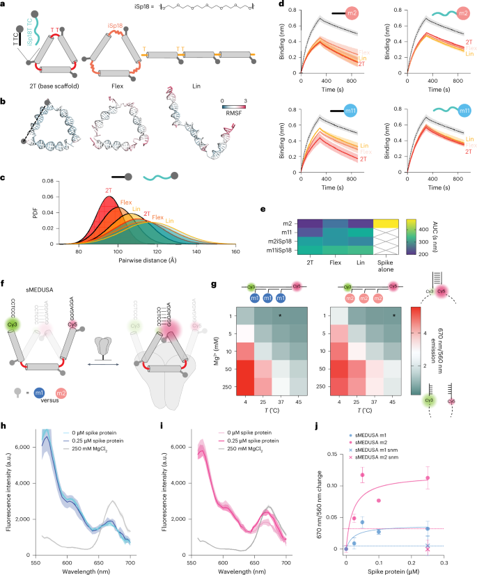
We produced a set of alternative structures with increased conformational flexibility (Fig. 5a, top) to explore how MEDUSA scaffold and linker configurations impact performance. The original cssDNA scaffold (2T) was linearized (Lin) to explore geometry and we replaced one of the T nucleotides by hexaethylene glycol (iSp18) in each of the three vertices to explore flexibility (Flex) (Fig. 5a). Increased flexibility of these alternative MEDUSAs was confirmed with OxDNA simulations49 (Fig. 5b,c and Supplementary Fig. 23). Local binding unit flexibility was modified by placing iSP18 linkers between the m2 and m11 binding units and their scaffold-hybridization regions. The inhibitory potential of all new scaffold and linker versions (Supplementary Fig. 24) was tested in a BLI competition assay with ACE2 dimer. Our results revealed that modifying the original geometry and flexibility of ligand presentation diminishes the performance of m2 and m11 assemblies. The most notable decrease was observed for assemblies with excessive local linker flexibility (Fig. 5d,e), regardless of the scaffold variant. The increase in the assembly’s core flexibility correlated with the decrease of the assembly’s inhibitory potential, which was especially prominent for the assemblies of the m11 binding unit, which was shown to gain most from multivalent presentation. We conclude that conformationally constrained scaffolds with target-specific geometry are crucial for the inhibitory potential of MEDUSA.
a, A diagram depicting the base multimerization scaffold (2T), its linear (Lin) variant and a variant with more flexible vertex regions achieved by substituting one of the T nucleotides with a hexaethylene glycol spacer (Flex). FNAPs with extended spacers separate the binding region from the assembly’s core. Corresponding coarse-grained models of the scaffold structures are given below. b, Close-to-average oxDNA models of the designed scaffolds. RMSF, root mean square fluctuation. c, Distribution of end-to-end distances between FNAP attachment points for various assemblies, based on coarse-grained simulations. The average distribution of all three distances is plotted for each particle. d, Competition BLI sensorgrams for FNAP assemblies with various scaffold and binding unit compositions; spike protein without assemblies is plotted with a dashed line. All measurements were performed in duplicate (n = 2, technical replicates), and average signals are plotted with s.d. ranges highlighted. e, A heatmap summarizing the mean areas under the curve (AUC) calculated from the BLI data for each scaffold and FNAP combination. f, Schematic of sMEDUSA, which features two conformational states in dynamic equilibrium, with the closed conformation being stabilized upon multivalent cis-interaction with the spike protein (shown in grey). g, Conformational state diagram for m1 and m2 devices as a function of temperature and Mg2+ concentration. The condition used in the spike protein binding assays is indicated by an asterisk. h, Fluorescence intensity spectral scans for m1 sMEDUSA. i, Fluorescence intensity spectral scans for m2 sMEDUSA. The respective spectral scans of sMEDUSAs in the presence of 250 mM MgCl2 are included as a reference for maximal FRET. j, Change in 670 nm/560 nm fluorescence ratio relative to sMEDUSA in buffer for m1 and m2 sMEDUSAs in the presence of increasing concentrations of spike protein, and for scrambled non-modified (snm) sMEDUSAs at 0.25 μM spike concentration. Dashed lines represent 670 nm/560 nm ratio change for m1 and m2 sMEDUSAs in the presence of 100 μg ml−1 BSA. All FRET assays were performed in triplicate (n = 3, technical replicates), with the average values plotted and s.d. ranges highlighted.
In an attempt to translate this optimal geometric binding and target selectivity into biotechnology tools, we explored if multivalent target-induced sensors could be generated from MEDUSAs. The conformation that facilitates the most effective ligand positioning should be favoured upon the multivalent engagement of its binding units with a multimeric target. Crucially, this only holds true if the binding units can engage in synergistic binding in cis. We devised a switchable MEDUSA variant (sMEDUSA) that can adopt two discrete conformational states: a suboptimal open state for multivalent cis-binding, and a more optimal conformationally constrained closed state. In the open state, sMEDUSA resembles the Lin structure, while in the closed state it is akin to the original MEDUSA. The structural basis for the switchable behaviour of sMEDUSA arises from its scaffold, which contains short (6-nt) complementary regions at the 5′ and 3′ termini flanking three FNAP-binding sites (Supplementary Fig. 25). Upon the hybridization of this scaffold with three binding units, a molecular switchable system with dynamic equilibrium between open and closed states is obtained. By introducing a Förster resonance energy transfer (FRET) pair at the root of the dynamic hairpin, the proportion of closed-state molecules can be monitored in bulk by ratiometric readout of fluorescence intensities at 670 nm (indicative of the closed conformation) and 560 nm (indicative of the open conformation) (Fig. 5f). We successfully validated the system by modulating the equilibrium between open and closed states using temperature and Mg2+ concentration: both a decrease in temperature and an increase in Mg2+ concentration led to the stabilization of the hairpin and a subsequent increase in the closed-state population (Fig. 5g).
To explore the dynamic range of the system and to test whether the same shift in the equilibrium between the open and closed conformations of sMEDUSA could be achieved by cooperative multivalent binding in cis, we conducted a sandbox experiment with sMEDUSAs featuring short DNA sequences with different GC content in place of FNAP-binding units (Supplementary Fig. 26). We observed that the conformationally constrained 2T MEDUSA target was the most effective in mediating the FRET increase of the sMEDUSAs, and the increase in GC content of the binding units correlated with the increase in FRET (Supplementary Fig. 26e). Next, we tested whether the conformational dynamics of sMEDUSA could be altered by the spike protein (Supplementary Fig. 27). We compared FRET efficiencies of m1 and m2 sMEDUSAs, along with sMEDUSAs with scrambled non-modified control sequences. Excitingly, the addition of the protein to the m2 sMEDUSA led to a more substantial increase in FRET compared with m1 sMEDUSA (Fig. 5h–j), indicating effective multivalent cis-binding to the spike protein and consequently a shift of the conformational state equilibrium towards the closed conformation. These data lay the foundation for the potential of the sMEDUSA system in molecular sensing applications as well as non-FRET-based sensor technologies.

